- Fully automated measurement process; 150 tests/hour throughput
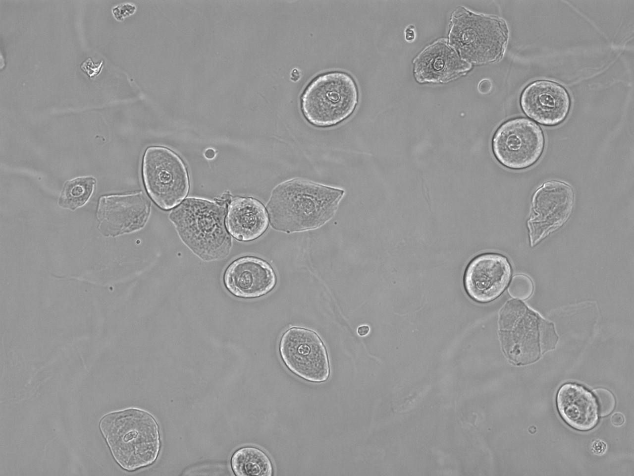
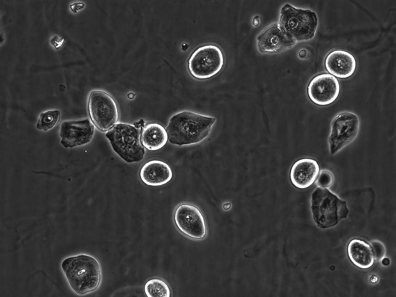
- Revolutionary particle visualization and recognition utilizing both bright-field and phase contrast microscopy
- Zoomable HPF-like images
- Live view mode: Real-time view of any viewfield of the cuvette to see moving microorganisms as well
- Dual-view for both bright-field and phase contrast images
- The only consumable is the UriSed Cuvette
- Improved consumable traceability: RFID based cuvette and rack identification
- Fully automated sample preparation requiring only low sample volume
- Automated QC analysis and maintenance procedures
- No need for liquid reagents or calibrator
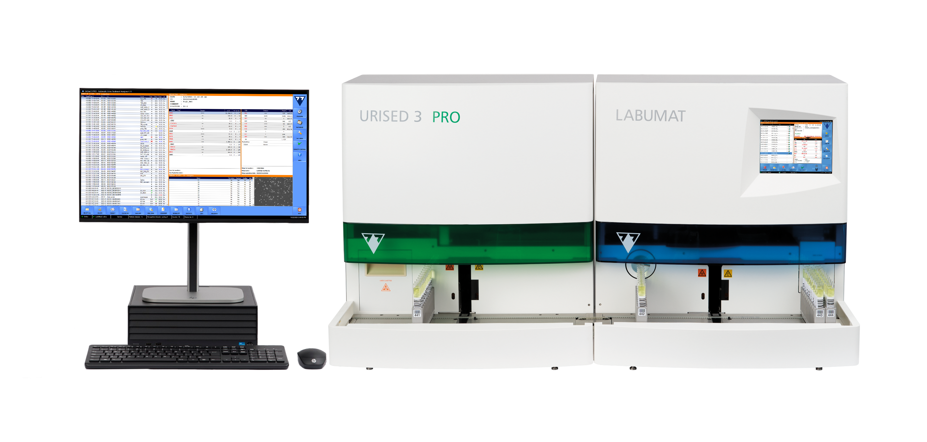
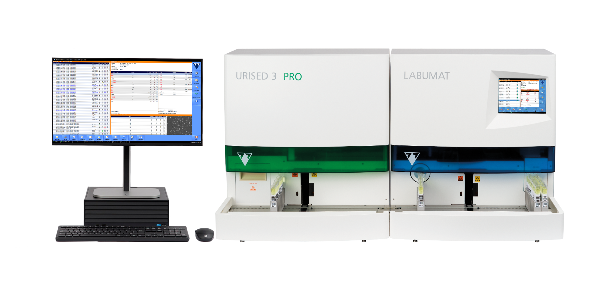
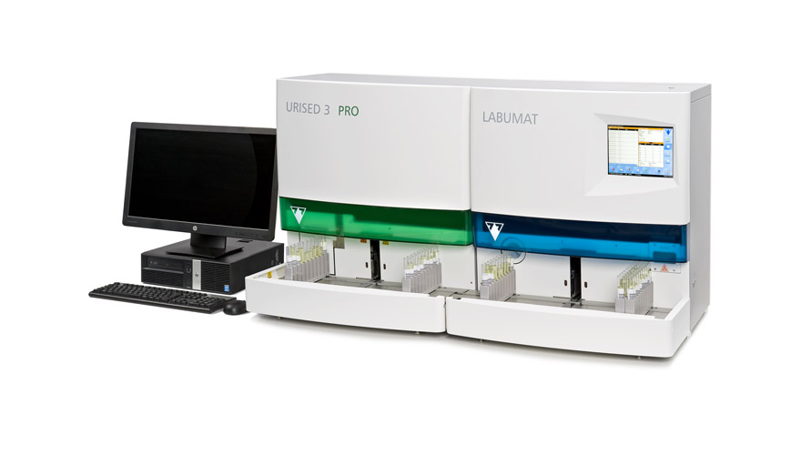
- UriSed 3 PRO and LabUMat 2 together make a Complete Urine Laboratory System
- Integration to laboratory or hospital information systems
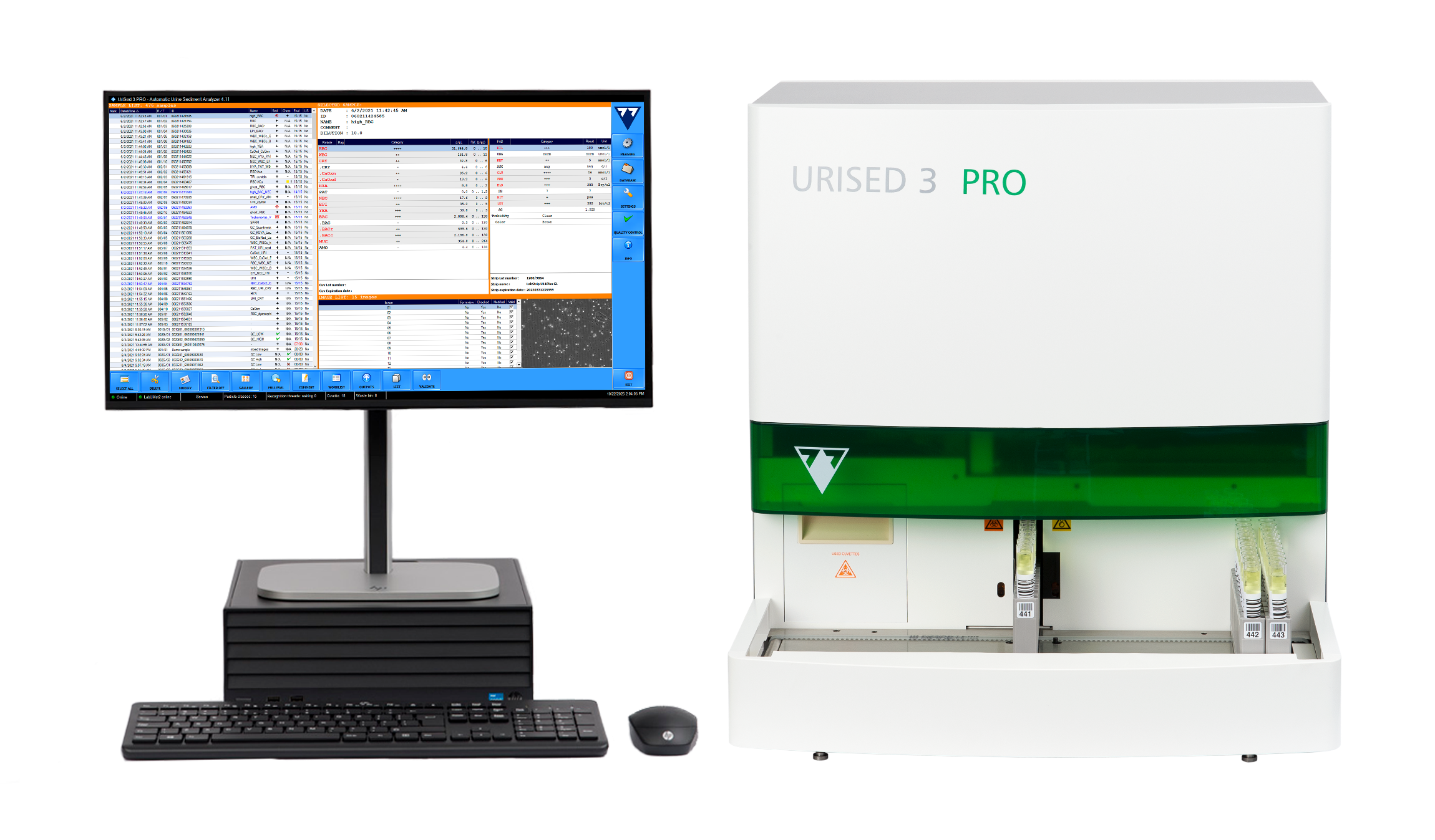
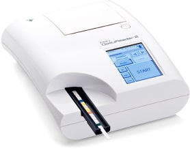
This instrument belongs to the Facelift generation of the UriSed technology. Its operation is based on the same patented measurement technique, which is actually the automation of traditional manual microscopy.
Working without any special liquid reagents UriSed 3 PRO performs sample preparation, takes multiple images of each sample through a built-in microscope, and evaluates the images using the Artificial Intelligence-based Evaluation Module (AIEM), a high-quality image processing software.
The UriSed 3 PRO microscopic urine sediment analyzer is a stand-alone instrument, which can be connected to the LabUMat 2 urine analyzer. Together, the two instruments make a Complete Urine Laboratory System.
Highlights
- Revolutionary particle visualizaton and recognition utilizing both bright-field and phase contrast microscopy.
- Fast and reliable instrument for walk-away operation. UriSed provides a reproducible method for the preparation and evaluation of urine samples,which is based on the automation of traditional microscopic investigation of urine.
- No special liquid reagent for sample evaluation. The cuvettes are the only consumable needed, and standard distilled water is the only solution required for operation.
- No carry over between samples because each sample is analyzed in a separate cuvette and because of the unique pipette washing technology.
- No obstruction or clogging caused by large sediment particles as the samples do not pass through any narrow channels inside the instrument during measurement.
- Low sample volume; minimum volume is checked by sample level sensor.
- Moderate centrifugation process that preserves the cells and casts in urine sediment so that they can be seen intact on UriSed Images.
- Zoomable HPF-like images
- Automatic and reliable image evaluation by the high-quality image processing software, AIEM.
- Manual microscopy mode. During the process the user has a live view of the cuvette.
- Quick particle option to improve manual marking of images.
- UriSed Technology provides whole view-field images through which no information can be lost; not only the automatically detected particles, but all details (e.g., rare types of urine particles, morphology) are visible.
- Education of laboratory staff or medical students through images displayed on full screen.
- Advanced result management, streamlined documentation through LIS, worklist handling.
- Automated QC analysis and maintenance procedures, software and language upgrades via USB stick.
- New auto-evaluation feature: Ghost RBC flag, RBC-Aca flag.
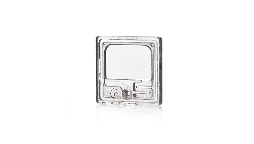
Urised Cuvette
The only consumable of the UriSed 3 PRO sediment analyzer is the UriSed Cuvette, which is a single-use high optical quality polycarbonate chamber.
Cuvettes are stored in containers of 50 pieces. An operator can load 12 containers (600 cuvettes) at once into the UriSed 3 PRO—enough to accommodate a typical day of sample anaylsis in most laboratories.
The analyzer automatically fills 200 µl of sample into the individual cuvettes. The filled cuvettes are centrifuged and forwarded to the microscope position where a built-in camera takes images of the settled urine particles.
Images & Results
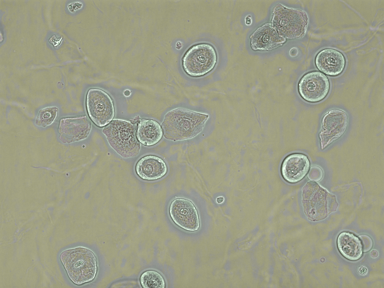
The truly unique advantage of UriSed Technology is, that it can provide not only images of individual particles, but also whole field of view images of the sample, as is visible under a microscope.
During the measurement process, UriSed 3 PRO produces three types of high-resolution images, a bright-field, a phase contrast and a composite microscopic image which is the combination of the former ones. The default setting is 15 images/sample, in which 2.2 µL of native urine is examined. Fifteen high-power field (HPF)-like UriSed 3 PRO images correspond to 10 standard manual microscopic images provided that a 400× magnification microscope is used and the concentration level of the sample is 20×, as suggested by the European Urinalysis Guideline.
UriSed 3 PRO images are evaluated by the AIEM, the automatic, real-time, image-processing software that scans the images and identifies urine sediment particles in them. The main advantage of this technology is that it uses an extremely wide range of information. By scanning the original whole field of view images the AIEM receives more input than that provided by the feature parameter-based methods of other technologies, thus providing a more accurate recognition algorithm.
All images can be accessed and displayed separately on screen from the user software. The recognized paricles are identified and labeled on the images by captions above them. The final result is generated by calculating quantitative concentration values and semi-quantitative category ranges for each sample. By allowing the operator to see the full context of the samples, rare particles that are not identified automatically by the AIEM can be marked manually quickly and easily.
Detecting and classifying the most frequently occurring urine sediment particles with specificities and sensitivities of >80% at high precision with no carryover, the UriSed 3 PRO fulfills the requirements of routine urine sedimentation analysis and offers an automatic and standardized alternative to manual microscopy.